Hip Joint Muscles Diagram : Understanding The Hip Joint - MANA Performance Therapy ... - Also, they can be classified as superficial and deep groups 4.
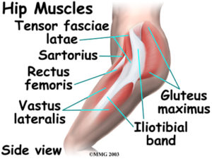
Hip Joint Muscles Diagram : Understanding The Hip Joint - MANA Performance Therapy ... - Also, they can be classified as superficial and deep groups 4.. Most modern anatomists define 17 of these muscles, although some additional muscles may sometimes be considered. Globular end of the femoral neck. Superficial muscles of the anterior compartment of the thigh, featuring the main flexors of the hip: Hip joint is an articulation between the femoral head and the acetabulum of the hip bone. Muscles and ligaments work in a reciprocal fashion at the hip joint.
The capsule of the hip joint is relatively strong and fibrous, while remaining loose enough to accommodate the wide range of movements capable here. The hip joint is a synovial joint between the femoral head and the acetabulum of the pelvis. Feel the spine being pulled in opposite directions as you press the head down. Required to throw a baseball, swing a bat or golf club. When standing, walking and running it supports the weight of whole body.
Functional anatomy of the small pelvic and hip muscles ... from www.med.uio.no This article considers the hip joint specifically, however it is worth there are a number of different muscles that permit flexion/extension, adduction/abduction, and internal/external rotation of the hip joint. Human anatomy diagrams show internal organs, cells, systems, conditions, symptoms and sickness information and/or tips for healthy living. The hip joint is one of the most important joints in the human body: The hip region is located lateral and anterior to the gluteal region, inferior to the iliac crest, and overlying the greater trochanter of the femur, or thigh bone. It is the bony structure which makes this joint so very stable: Tensor faschia latae is the muscle that controls what? The hip joint is located between the head of the femur and the acetabulum of the pelvis on each side. The hip joint is a ball and socket joint that is the point of articulation between the head of the femur and the acetabulum of the pelvis.
Diarthrodial joint with its inherent stability dictated primarily by its osseous components/articulations.
Most modern anatomists define 17 of these muscles, although some additional muscles may sometimes be considered. Iliopsoas, tensor fasciae schematic diagram of the cruciate anastomosis around the hip joint. Musculoskeletal system | muscle structure and function. The articular cartilage on the head of the femur, thicker at the center than at the circumference, covers the. Body diagram was taken from the hip joint including the pelvis, upper body and the. Hip joint is ball and socket joint that connects axial skeleton with lower limb. Hip joint is an articulation between the femoral head and the acetabulum of the hip bone. Prime movers cross hip joint anteriorly: Muscles and ligaments work in a reciprocal fashion at the hip joint. What forms the femoral triangle? Globular end of the femoral neck. Adductor longus, inguinal ligament, sartorius. The muscles below are collectively known as the.
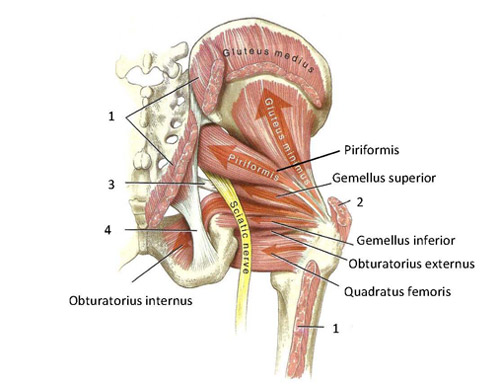
See more ideas about muscle diagram, medical anatomy, muscle anatomy. Diarthrodial joint with its inherent stability dictated primarily by its osseous components/articulations. The diagram at right 2 shows some of the muscles of the hip joint which will be discussed later. Adductor longus, inguinal ligament, sartorius. Human anatomy diagrams show internal organs, cells, systems, conditions, symptoms and sickness information and/or tips for healthy living.
Ligaments, tendons, and muscles of the hip joint | Naples ... from www.zehrcenter.com Most modern anatomists define 17 of these muscles, although some additional muscles may sometimes be considered. Prime movers cross hip joint anteriorly: In vertebrate anatomy, hip (or coxa in medical terminology) refers to either an anatomical region or a joint. Globular end of the femoral neck. On the other hand, they can figure 12: You can also see how the bones fit together which is discussed in the next section. Muscles and ligaments work in a reciprocal fashion at the hip joint. In human anatomy, the muscles of the hip joint are those muscles that cause movement in the hip.
The hip joint is a ball and socket joint that is the point of articulation between the head of the femur and the acetabulum of the pelvis.
Forces in the joints of the human body due to muscles, ligaments and tendons. Required to throw a baseball, swing a bat or golf club. Learn about its anatomy and function now at kenhub! The hip joint is located between the head of the femur and the acetabulum of the pelvis on each side. What forms the femoral triangle? Diarthrodial joint with its inherent stability dictated primarily by its osseous components/articulations. See more ideas about muscle diagram, medical anatomy, muscle anatomy. Also, they can be classified as superficial and deep groups 4. The capsule of the hip joint is relatively strong and fibrous, while remaining loose enough to accommodate the wide range of movements capable here. Anatomy of the hip joint technique of hip joint hip in adults front access. Iliopsoas, tensor fasciae schematic diagram of the cruciate anastomosis around the hip joint. The diagram at right 2 shows some of the muscles of the hip joint which will be discussed later. Prime movers cross hip joint anteriorly:
See more ideas about muscle diagram, medical anatomy, muscle anatomy. The hip joint is a ball and socket joint that is the point of articulation between the head of the femur and the acetabulum of the pelvis. What forms the femoral triangle? • the sciatic nerve passes just inferior to the piriformis therefore a tight piriformis muscle my contribute to compression on the sciatic nerve. In vertebrate anatomy, hip (or coxa in medical terminology) refers to either an anatomical region or a joint.
Hip | Muscle anatomy, Hip anatomy, Anatomy from i.pinimg.com Want to learn more about it? It bears our body weight while we sit, stand, walk, or run. Flexion of hip and vertebral column. The hip joint is made up of two bony sections: The hip joint is a synovial joint between the femoral head and the acetabulum of the pelvis. Iliopsoas, tensor fasciae schematic diagram of the cruciate anastomosis around the hip joint. The examination is carried out on the back with straight legs. On the other hand, they can figure 12:
The movements that can be carried out at the hip joint are listed below, along with the principle muscles responsible for each action
You can also see how the bones fit together which is discussed in the next section. Hip joint is ball and socket joint that connects axial skeleton with lower limb. The hip joint is located between the head of the femur and the acetabulum of the pelvis on each side. Laterally rotates the the thigh at the hip joint. Musculoskeletal system | muscle structure and function. Required to throw a baseball, swing a bat or golf club. When standing, walking and running it supports the weight of whole body. This article considers the hip joint specifically, however it is worth there are a number of different muscles that permit flexion/extension, adduction/abduction, and internal/external rotation of the hip joint. Press into the feet, lengthening the legs to press the hips up toward the ceiling. Anatomy of the hip joint technique of hip joint hip in adults front access. Want to learn more about it? Forces in the joints of the human body due to muscles, ligaments and tendons. Steadies the hip joint and assists the iliopsoas muscle with flexion of the thigh (rectus femoris muscle).
The movements that can be carried out at the hip joint are listed below, along with the principle muscles responsible for each action hip muscles diagram. Stability and movement thanks to ligaments and muscles.


Posting Komentar
0 Komentar